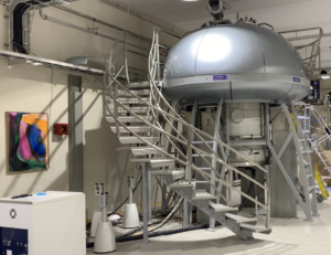
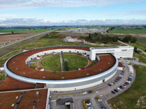
Lectures in the integrative structural biology course.
Learning outcomes
After the course the participants will be able to:
1.… describe the sample requirements for and interpret the nature of structural information obtained from the techniques:
a. Macromolecular X-ray crystallography
b. Macromolecular neutron crystallography
c. Small angle X-ray scattering
d. Small angle Neutron scattering
e. Single particle Electron Microscopy
f. Macromolecular NMR spectroscopy
2.… explain how information from the different structural biology techniques can be combined to resolve biological questions
3.… recognize how computational chemistry techniques can be used to combine data from multiple structural biology techniques
Lecture outline examples
Macromolecular nuclear magnetic resonance spectroscopy (NMR)

NMR is a technique where the fundamental concept is to detect signals from individual atoms, giving insight on the atomic/molecular level. The strength in NMR lies in being a complementary technique to Cryo-Em and Crystallography once the structure is known at atomic, or near atomic level. NMR can be used to study the interactions in protein complexes, between a protein and a small molecule or other interaction partner. Dynamics of proteins are commonly studied using NMR.
NMR is often applied in studies where questions about function of proteins are in focus, taking advantage of the fact that the protein is in solution and can easily be titrated with possible ligands. Finding new biding molecules that could be used as starting point for drug development can be achieved using screening techniques where a library of roughly 1000 compounds can be analyzed in 1-2 days. NMR studies rely on the protein being in solution and within a certain size range, but the range can, however, be extended by proper isotope labeling to cover sizes from peptides all the way up to 100kD.
The practical part will cover how to determine Kd for a protein/ligand complex, using data from an NMR titration series.
We will work in the Software ccpn v3 that will be available through common source.
After this course, you will be able to known how you should label your protein optimally to study your scientific question, have insight into who to contact for support in the process, and have gone through the practical steps of determining the dissociation constant for a protein/ligand complex.
Crystallography

Crystallography is a scattering technique that makes use of the signal amplification in crystals. Having many molecules ordered in precisely the same orientation allows atomic level information to be extracted from the inherently weak scattering of X-rays or neutrons. To compensate for the weak scattering large facilities are needed to create brilliant beams of X-rays or neutrons. While X-ray crystallography is a workhorse technique for protein structure determination, neutron crystallography can provide unique information about hydrogen atoms.
X-ray crystallography is often the technique of choice for protein structure determination, but the ability to grow well-ordered crystals is often the bottleneck. Highly automated approaches are used to find the right crystallization conditions and the X-ray data collection is also automated for high throughput. While X-ray crystallography can determine the protein structure rapidly and accurately, hydrogen atoms are generally invisible. Neutron crystallography can be used to determine the hydrogen positions, but due to the weak neutron sources available much larger crystals are needed. Optimising the crystal growth for neutron crystallography is a major task.
In the practical we will focus on how to interpret the density maps from both X-ray and neutron data from a fatty acid binding protein.
We will use software from the PHENIX package as well as Coot that will be available through common source.
After this course you will understand the principles of crystallographic structure determination and know what the benefits and limitations of X-ray and neutron crystallography are. You will also know whom to contact in Sweden for support on X-ray and neutron crystallography.
Two examples of papers to read about what we will address in the practical are:
Overview of macromolecular X-ray crystallography
A beginner’s guide to neutron macromolecular crystallography and references therein
Structural mass spectrometry, XL-MS and HDX-MS
Hydrogen-deuterium exchange mass spectrometry (HDX-MS) and crosslinking mass spectrometry (XL-MS) are both techniques used to study the structures and interactions of proteins or protein complexes in solution. HDX-MS can measure the dynamics and interactions between proteins and ligands by hydrogen deuterium exchange, whereas XL-MS is based on covalently crosslinking the proteins. The techniques are different but complementary to each other, and are both based on measuring information on the peptide level.
HDX-MS and XL-MS have different abilities and limitations. HDX-MS works in solution with only a few (up to three) purified components, can detect dynamic changes in structure and interaction interfaces, and is complementary to several other structural biology techniques, such as crystallography and cryoEM. XL-MS can be done on several different sample types ranging from intact cells in culture to purified proteins in solution, and gives distance constraints between two peptides, which can be used in modeling or to complement data from NMR, crystallography or cryoEM. Even though both HDX-MS and XL-MS work on the peptide level, the resolution of HDX-MS is higher, and can at its best pinpoint events on the individual amino acid level.
HDX-MS has recently been extensively applied in studies of the spike protein for SARS-COV-2, i.e. for epitope mapping to find potential neutralizing antibodies. In the practical session we will use the software HDexaminer or Masspecstudio through/via PreSTO to analyze data from a previous publication ( https://www.nature.com/articles/s41592-019-0459-y).
After this course you will be able to decide what structural mass spectrometry can be used for, whether these approaches are suitable to study your scientific question, have insight into who to contact for support in the process, and have gone through the practical steps of analyzing HDX-MS data. XL-MS data analysis will also be demonstrated during the lectures.
You can read more about structural mass spectrometry here:
Cryo-EM single particle analysis (SPA)

Cryo-EM SPA allows us to determine high-resolution structures of protein, protein-protein and protein-oligonucleotide complexes starting from transmission electron microscopy pictures of many hundred thousand purified molecules trapped in a thin layer of vitrified ice. The pictures are computationally analyzed and sorted in different classes based on their orientation on ice and their similarities in general. Classification can be performed both in 2D and 3D so that different conformations of the same macromolecular complex can be separated and resolved, and molecular movements inferred.
Cryo-EM works best for specimens larger than 100kDa in size even though proteins as small as 50kDa can also be analyzed. Achievable resolution can be better than 2 Å, but is generally between 2 and 4 Å. Moreover, the resolution in cryo-EM can vary enormously within the same structure depending on domain flexibility or occupancy. It is common to have less resolved regions that can be interpreted using a combination with other techniques such as X-ray crystallography and NMR, or model prediction methods such as AlphaFold2. The combination of cryo-EM with molecular-dynamics simulations can also be extremely powerful.
Another application of cryo-EM is cryo-electron tomography (cryo-ET), used for the analysis of macromolecules in their cellular environment or for the study of pleiomorphic specimen in general (e.g. full non-regular viruses). Another application is cryo-electron diffraction of nano crystals, commonly known as micro-ED, which is strongly related to X-ray crystallography but uses electrons as the medium for diffraction. We will however not touch on these two techniques in this course.
In the practical part, we will perform a simple pipeline of cryo-EM single particle analysis from raw image analysis to 3D refinement and model fitting. After this course, you will be able to known how you should approach your first cryo-EM project, what are the most important steps in obtaining a final structure and how to interpret different parts of the determined cryo-EM map.
If you are interested in reading more about cryo-EM and its coupling with other techniques and fields in structural biology here are some links to online courses and reviews.
https://cryo-em-course.caltech.edu/
Small Angel Scattering (SAS)

Scattering techniques provide information on atomic or molecular scale structure. The techniques used for scattering include X-ray crystallography, powder diffraction and small-angle scattering.
Small-angle X-ray scattering (SAXS) can provide information on the inner structure of disordered or partially ordered materials that are challenging to crystallize. Often used in soft matter research it is an ideal technique for the study of structures in the nanometer to micrometer range. The method can provide information on the averaged particle size, shape, and distribution, as well as surface-to-volume ratio. Small-angle scattering can for example be used to observe protein structure and dynamics in solution, specifically conformational changes upon external stimuli. Several instruments at MAX IV offer SAXS and two instruments, CoSAXS and ForMAX, are dedicated for this technique.
CoSAXS
The CoSAXS beamline is a state-of-the-art multipurpose small-angle X-ray scattering instrument that can be used to probe the structure of proteins and other biological macromolecules in solution and where the experimental parameters can easily be varied. Apart from biomolecules SAXS can be used to study colloidal systems, polymers, food, thin films such as for example from mucous membranes. It is also an optimal technique for studying formulations of for example drug delivery system.
ForMAX
Many biological and soft-matter systems exhibit structural hierarchy, i.e., they show important structure at many different length scales. In order to understand the structure-function relationship in such materials, multiscale structural characterization is needed. The beamline ForMAX will address this need by combining two complementary techniques – full-field imaging for microscopic characterization as well as small- and wide-angle x-ray scattering (SWAXS) for nanoscopic characterization. A combination of these techniques, scanning SWAXS imaging, will provide unique access to the full range of micro- and nanoscopic length scales. This will be useful for multiscale structural characterization of a wide range of biological and soft-matter materials, with a special focus on bio-based fibrous materials. The temporal resolution will allow in-situ studies of many processes.
To understand the application of SAXS to protein structure we will use a program suite for small-angle scattering data analysis from biological macromolecules on a model protein. The Data analysis software suite (ATSAS 3.0.5) has been developed by the EMBL Hamburg and can be downloaded via: https://www.embl-hamburg.de/biosaxs/software.html